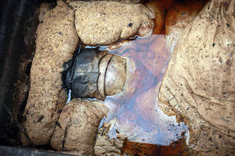
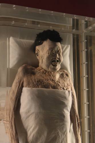
Strannik-57 писал(а):И ещё одна непонятка из этой серии : китайские мумии в саркофагах....Аутолиза, судя по фото, не наблюдается. Пусть даже и с датировками наврали.
)))) Это 20 век. Мумия династии Мин в ботах фирмы Зингер
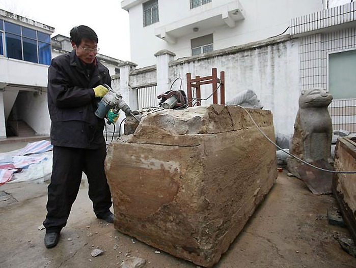
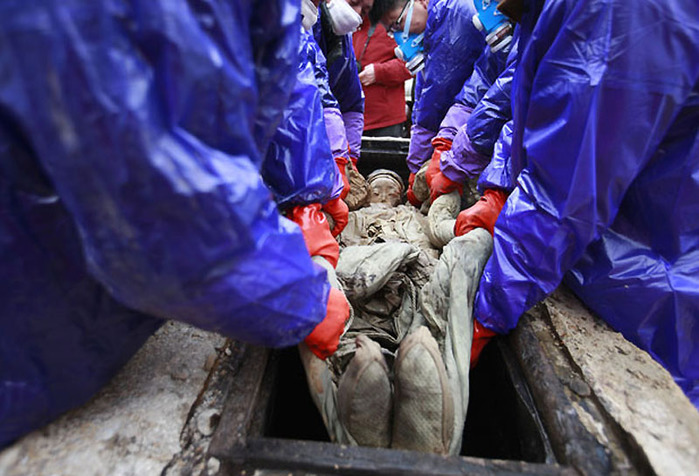
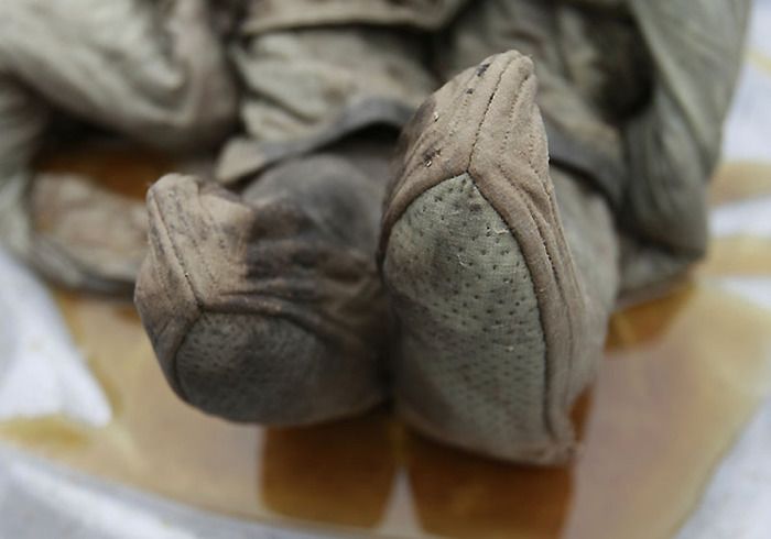
Strannik-57 писал(а):Саркофаг таки ящик. А для чего ящик - это дело вкуса. Для засолки огурцов или выгонки первача. Но в свете Вашего утверждения о "литье" саркофагов, перечисление таких пород как: базальт; гранит; мрамор...э....смущает. Вы уверены, что это ГРАНИТНОЕ именно литьё? А если таки литьё, то таки НЕ ГРАНИТНО-БАЗАЛЬТОВО-МРАМОРНОЕ ? А если даже это природный выдолбленный гранит-базальт, то как долго в нём сохранится влага от аутолиза тела массой 100 кг.? Ведь даже эти материалы являются водопроницаемыми.
А чего вам не нравится? Его и
в Барнауле льют.
Литье оно и в Египте литье.
Вот еще
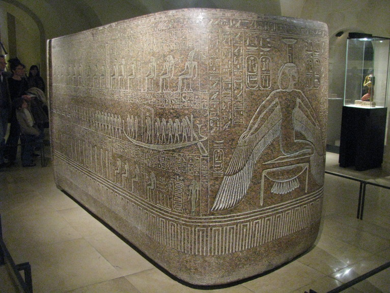
Strannik-57 писал(а):А попробуйте эту. Может это покажется Вам более информативным.
Ссылка
Хорошо работаете, молодца!
Реферат работы, присланной "палеонтологом", с автографами представителей 2 музеев и института Карнеги, с обратным адресом в Киле (Германия) ))))
Читайте
это
"Открытие", о котором заявлено в 1993, давно опровергнуто, извлечено на свет в "нулевых" и гуляет само по себе.
Не рискую приводить свой английский (я его "со словарем"), просто суть: бактериальные микропленки, современного происхождения, с контрольными проращивания аналогов и т.д.
Свернутый текстA scanning electron microscope survey was initiated to determine if the previously reported findings of “dinosaurian soft tissues” could be identified in situ within the bones. The results obtained allowed a reinterpretation of the formation and preservation of several types of these “tissues” and their content. Mineralized and non-mineralized coatings were found extensively in the porous trabecular bone of a variety of dinosaur and mammal species across time. They represent bacterial biofilms common throughout nature. Biofilms form endocasts and once dissolved out of the bone, mimic real blood vessels and osteocytes. Bridged trails observed in biofilms indicate that a previously viscous film was populated with swimming bacteria. Carbon dating of the film points to its relatively modern origin. A comparison of infrared spectra of modern biofilms with modern collagen and fossil bone coatings suggests that modern biofilms share a closer molecular make-up than modern collagen to the coatings from fossil bones. Blood cell size iron-oxygen spheres found in the vessels were identified as an oxidized form of formerly pyritic framboids. Our observations appeal to a more conservative explanation for the structures found preserved in fossil bone.
Go to:
Introduction
The previous discovery of soft, pliable tissues recovered from the dissolved remains of Tyrannosaur bone in 2005 [1], potentially marked a major turning point in the science of paleontology given that it extended the known range of preserved biomolecules by many orders of magnitude. The implication that these were preserved dinosaurian soft tissues held the promise of biologic investigations of extinct animals. The original discovery centered on several tyrannosaur specimens. From this single report, it could not be determined if this was a wholly unique one-of-a-kind preservation, or these structures remained undiscovered in other fossil material. Subsequent investigations [2] showed that these microstructures existed across a range of time and taxa and ruled out a one-time exceptional preservation. The previous work required that the fossil bone be dissolved in acid to expose the preserved microstructures.
A new line of investigation was undertaken to detect the material in unaltered bone. Furthermore these structures should be occurring commonly in bone from the same formations. This work expands on these initial investigations by examining the interior of the fossil dinosaur bone prior to dissolution in acid using scanning electron microscopy (SEM) and energy dispersive spectroscopy (EDS). The survey totaled more than 200 hours of SEM time, covered seven geologic formations and more than fifteen genera outlined in Table 1. The data and findings presented identify a bacterial biofilm that mimics soft structures previously thought to be biological tissue. This explanation is in marked contrast to the concept of an exceptional preservation scenario of dinosaurian soft tissue and represents a plausible alternative hypothesis.
Table 1
Table 1
Specimens examined in survey.
Four categories of tissues were initially discovered in 2005 [1]: (A) Clusters of spheres that showed an iron-oxygen elemental signature appeared red under the light microscope. (B) Soft, branching, tube-like structures that contained spheres. (C) Free floating “osteocytes” complete with fillapodia and (D) a filamentous mass that remained pliable and elastic. Subsequent tests using immunochemistry showed positive for proteins [3]. Three of these structures were found commonly in this survey and discussed below.
Go to:
Materials and Methods
Scanning electron microscopes used were JEOL T300, Zeiss Supra and Cambridge S200. Light microscopes were Zeiss Axiomat, Nikon SMZ-U and Unitron. EDS spectra were taken with Kevex Delta 5, EDAX Apollo 40 running under EDAX Genesis/Pegasus and Link LZ5 running under WinEDS.
SEM specimens were prepared by pressure fracturing and selected pieces approximately 10 mm square were fixed to aluminum stubs with high purity carbon tabs. Initial specimens were uncoated to minimize potential disruption of internal contents. Subsequent specimens were gold coated with a Bio-Rad E5000 sputter coater or Denton Desk IV turbo coater with Pd or Pt target. Low dot-pitch element maps were run at 15 kV. The maps of individual elements were combined with a high-resolution secondary electron images to produce a high resolution colored element maps.
Modern biofilms were grown with the following method. Two gallons of water obtained from a local pond was placed in a new five gallon bucket and recirculated with a small pump at room temperature. Five new microscope slides were placed in the bottom with 100 mg of glucose nutrient added to the water. Water samples were taken every two days to monitor microbe population. One slide was removed and examined under the light microscope every few days to monitor biofilm accumulation rate. Slides were allowed to desiccate at room temperature over several days. Microbial communities could clearly be seen in the hydrated biofilms under the light microscope but subsequent examination under SEM showed only a smooth undulating profile.
Fourier Transform Infrared Spectroscopy (FT-IR) was used to investigate the specimens' molecular structure. A turtle carapace from the Hell Creek formation was selected for spectroscopy because of its proportionally large chambers in the trabecular bone that allowed scraping the coatings loose. Two milligrams of material was ground with 450 milligrams of potassium bromide (KBr) and pressed into a pellet using 8 tons pressure. Modern biofilms grown on microscope slides in pond water were allowed to desiccate for 7 days and 2.5 milligrams were pressed into a KBr pellet as above. A 2.5 milligram sample of desiccated tendon from a chicken was ground with KBr and pelletized. Spectrums were taken on a Nicolet 510P bench at 1 cm−1 resolution with a minimum of 15 scans. Infrared flux was matched within 5% for all specimens and a clean KBr pellet used for background subtraction between specimens. Excel cross correlation routines were used to determine percentage of similarity for spectrums.
Framboids were individually extracted from fractured dinosaur bone fragments with a magnet and transferred to a carbon sticky tab on an SEM stub. EDS was performed on a small section of the sphere at 20 kV in the area shown with the spectrum in figure 1.
Figure 1
Figure 1
EDS spectrum of framboid.
Multiple specimens were pressure fractured and 10–20 mm fragments selected for demineralization in 0.5 M ethylenediaminetetraacetic acid (EDTA) (pH 8.0) in individual plastic containers at room temperature. Resident times ranged from several days to several weeks depending on specimen resistance. Baths were changed at approximately three day intervals with fresh acid. Remaining structures were either photographed directly in the baths at low magnification 7–75× or removed for higher power imaging.
All specimens for carbon dating were handled under a flow hood with clean sterile gloves and instruments. The specimens were pressure fractured to reveal fresh surfaces. A bone fragment from the Lance formation was microscopically examined and coatings that appeared to have been dislodged were removed for analysis. Fifty milligrams of material were sent to Geochron Labs, Cambridge Mass. for accelerator mass spectrometry (AMS) analysis. The results were 139.01%±0.65 of modern (1950) of 14C activity.
Go to:
Results
The hollow morphology of the tyrannosaur femur supported the general idea that an exceptionally well-preserved bone may act as a containment vessel for biomolecules. To test this concept, a perfectly preserved turtle phalange showing no cracks or deformities (Fig. 2) was first selected for SEM examination. The specimen was pressure fractured and directly examined uncoated in the SEM. A cluster of spheres approximately 10 microns in diameter, similar to Fig. 3, was discovered almost immediately. Subsequent EDS showed an iron-oxygen signature. Continuing SEM surveys of multiple specimens from the Lance, Hell Creek, Chadron and Pierre Shale formations all showed similar iron-oxygen spheres ranging in size from 5–29 microns. Examination of demineralized specimens under the light microscope displayed small red spheres clustered in the tubular structures (Fig. 4A). Discovery of these spheres in an ammonite suture indicated they had no relationship to iron derived from blood. These spheres were identified as framboids which are seen world wide in black smokers, algal mats and are commonly found in sediments [4].
 А наличие такой лаборатории и группы исследователей, и самого такого исследования - никто не оспорил. Пройдясь к источникам, можно увидеть довольно много интересных фото объектов исследования. Довольно подробное описание процесса, результатов и выводов.Осторожные и сдержанные, но не менее от этого сенсационные,официальные заявления руководства института и проекта. Опять таки без оспаривания. Сомнений высказано много. Опровержений - ни одного.
 А если таки литьё, то таки НЕ ГРАНИТНО-БАЗАЛЬТОВО-МРАМОРНОЕ ? А если даже это природный выдолбленный гранит-базальт, то как долго в нём сохранится влага от аутолиза тела массой 100 кг.? Ведь даже эти материалы являются водопроницаемыми.
 По Вашему все портные должны быть криворукими? Старорусская вышивка "белым по белому" или златом, надо полагать, производилась исключительно современными навороченными агрегатами?
